Figure 1.
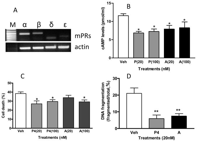
The mPRs are expressed in mouse neuronal GT1-7 cells (A) and low nanomolar concentrations of allopregnanolone mimic the inhibitory effects of progesterone on cAMP production (B), cell death (C) and DNA fragmentation (D) in neuronal GT1-7 cells. (A) Detection of mPRα, mPRβ, mPRδ and mPRε mRNAs by RT-PCR. (B) Cultured mouse neuronal GT1-7 cells were incubated overnight in serum-free media to reduce background adenylyl cyclase activity before treatment for 15 min. with vehicle, progesterone or allopregnanolone (20 nM and100 nM). The cAMP concentrations in cell lysates were measured with an EIA kit (Cayman, Ann Arbor, MI). (C,D) Cells, 70% confluent, were cultured for 4 days in serum-free media alone (vehicle) of media containing progesterone or allopreganolone. Approximately 500 cells were counted for cell death and DNA fragmentation after staining with trypan blue and by TUNEL assay, respectively, as described previously [45]. Veh: vehicle, P4: progesterone, A: allopregnanolone. N=6. * P<0.05, ** P<0.001 compared to vehicle controls (one-way ANOVA and Tukey’s ).

