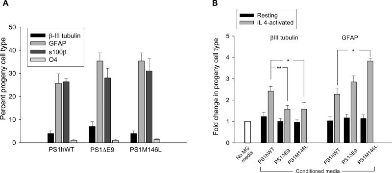
Figure 6. Differentiation pattern of cultured NPCs is modulated by FAD-linked PS1 mutant expressing microglia.
(A) Progeny cell types generated by NPCs in the multipotency assay were quantified by sampling a minimum of 300 cells as marked by DAPI or propidium iodide+ nuclei (mean ± SEM of the results obtained from independent cultures established from 6 animals per group).
(B) Fold changes in the number of βIII-tubulin+ neuronal or GFAP+ astrocyte progeny cell type derived from PS1hWT NPCs exposed to CM collected from resting or IL4-activated MG, over differentiated PS1hWT NPC cultures in the absence of MG CM (mean ± SEM; N = 6). The asterisk indicates significant difference from PS1hWT MG CM at * P < 0.05 and ** P < 0.01.

