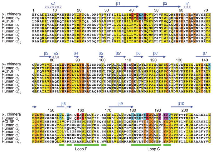
Figure 1.
Sequence and numbering of the α7–AChBP chimera and its alignment with related AChR sequences. Orange indicates invariant residues and yellow indicates partially conserved residues. Secondary structures are shown schematically above the sequences. Putative functionally important residues for ligand recognition (pink), signal transduction (blue) and inorganic ion binding (red) are shown. Loops F and C are indicated by green bars.

