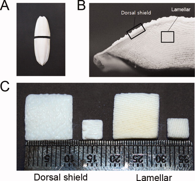
Figure 1.

Preparation of dorsal shield and lamellar blocks from cuttlefish bone. A: Whole cuttlefish bone in planar view and transverse section. B: A digital image of the transverse section through the cuttlefish bone. C: Prepared cuttlefish bone blocks (10 × 10 × 2 mm3 and 4 × 4 × 1 mm3) for cell experiments. [Color figure can be viewed in the online issue, which is available at wileyonlinelibrary.com.]
