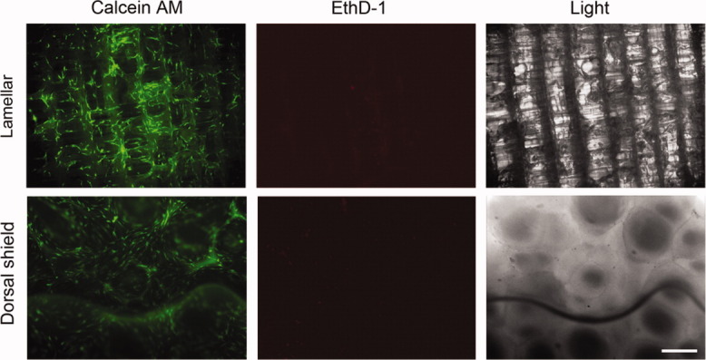
Figure 4.

Cells were seeded on lamellar and dorsal shield blocks of cuttlefish bone and cultured in normal growth media. After 3 days of culture, the viability/cytotoxicity assay was performed. Calcein acetoxymethyl (AM) stained healthy cells green and ethidium homodimer-1 (EthD-1) stained the nuclei of dead cells red. Light images show the surface morphology of each block. Scale bar represents 50 μm. [Color figure can be viewed in the online issue, which is available at wileyonlinelibrary.com.]
