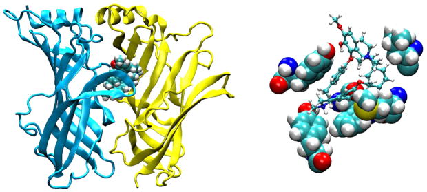
FIGURE 16.
Orientation of d-tubocurarine bound to AChBP (92). Left: principal subunit is yellow and the complementary subunit blue, while d-tubocurarine is shown as space-filling. Right: key contact residues are shown as space-filling (Y192, Y89, W143, M114, L112), while d-tubocurarine is shown as ball and stick.

