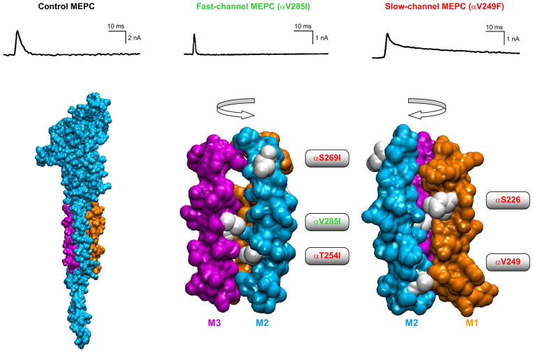
FIGURE 19.
Overview of congenital myasthenic syndromes due to mutations in AChR transmembrane domains. Top panel compares time courses of miniature endplate currents from control (black), fast channel (green), and slow channel (red) endplates [From Engel et al. (84)]. Bottom panel shows a homology model of the AChR α-subunit (based on PDB code 2BG9) as space-filling, with TMD2 highlighted in blue, TMD3 in magenta, and TMD1 in orange. Identified CMS mutations are indicated and shown with side chains white.

