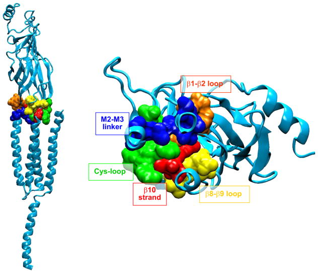
FIGURE 5.
Interface dividing ligand binding and pore domains of the α-subunit from a homology model of the human AChR generated using the Torpdeo AChR as a template (276). Consecutive residues are highlighted in a single color and labeled. In the left panel, the pore runs vertically along the left side, and in the right panel, the pore is just beneath the β1-β2 loop coming out of the plane of the page.

