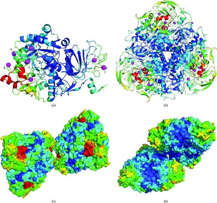
Figure 4.
Urease structure with residues colored according to their radiation sensitivity at 300 K. (a) The C chain, with magenta balls indicating positions of the peaks of the ‘ripples’ in Fig. 3 ▶. Five of the balls are near the active-site flap (red), while the other two are near a copy of the flap in the trimer-of-trimers unit (b). The B chain is both solvent-exposed and near the flap, which may explain its enhanced sensitivity at 300 K. (c) A crystal contact between two symmetry-related copies of the flap (each in a different copy of the biological unit) is shown. (d) A second crystal contact located on the other side of the trimer-of-trimers unit as the flap. These visualizations were created with PyMOL (http://www.pymol.org).

