
Fig. 2.
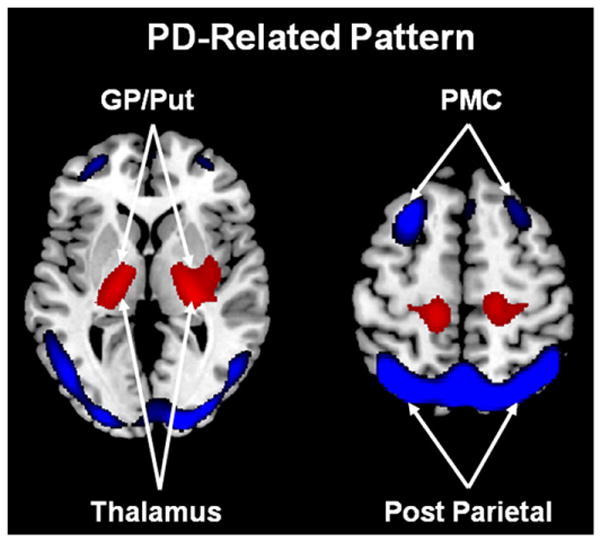
Abnormal metabolic motor networks in Parkinson’s disease. The PD-related motor pattern (PDRP) identified by spatial covariance analysis of FDG PET scans from 33 PD patients and 33 age-matched normal volunteer subjects. This pattern is characterized by relative hypermetabolism (red) in the globus pallidus/putamen (GP/Put), thalamus, pons, cerebellum, and sensorimotor cortex, associated with metabolic decreases (blue) in the lateral premotor cortex (PMC) and parieto-occipital association regions (Ma et al., 2007). Representative slices of the covariance map were overlaid on a standard MRI brain template.
From Eidelberg (2009) Fig. 1A.