
Fig. 5.
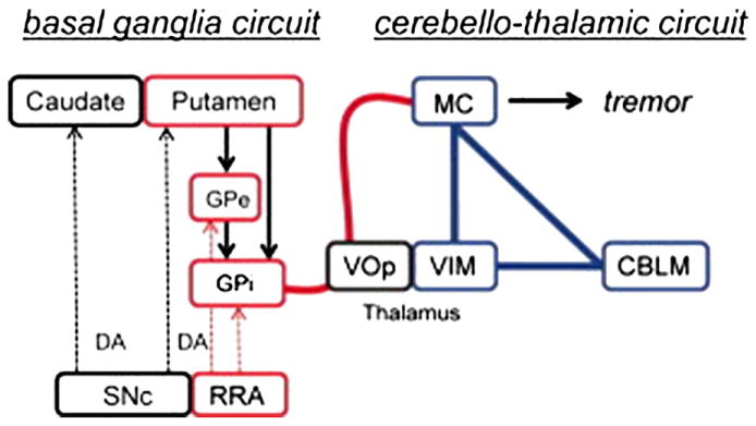
A model of cerebral mechanisms underlying Parkinson disease (PD) resting tremor proposed by Helmich et al. (2011). The authors propose that PD resting tremor emerges from the ventral intermediate nucleus of the thalamus (VIM)–motor cortex (MC)–cerebellum (CBLM) circuit (shown in blue), when triggered by transient pathological signals from the basal ganglia motor loop (shown in red). In tremor-dominant PD, the basal ganglia (globus pallidus internus [GPi], globus pallidus externus [GPe], and putamen) have increased connectivity with the VIM–MC–CBLM circuit through the MC (thick red line), and the basal ganglia are activated at critical time points in the tremor cycle. VOp = thalamic ventralis oralis posterior nucleus. DA = dopamine; SNc = substantia nigra pas compacta.
From Helmich et al. (2011) Fig. 4.