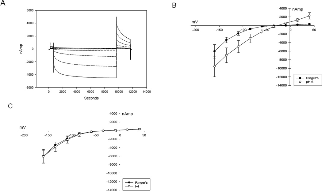
Figure 1.
A. Voltage step protocol demonstrating activation of current at each increasingly negative voltage step in a ClC-2 expressing oocyte. B. I – V plot of ClC-2 - βAR expressing oocyte showing enhanced activation with pH 6.0 superperfusate. C. I – V plot of ClC-2 and βAR expressing oocyte demonstrating no response to isoproterenol and IBMX.

