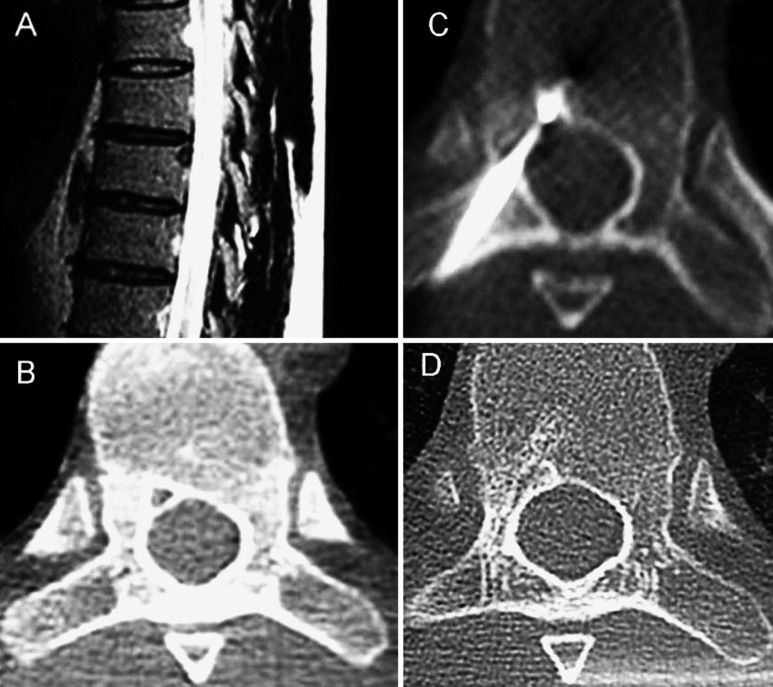
Fig. 1.
A The MRI of the thoracic spine on T2 weighted image sagittal slice, showing a 7-mm low T2 intensity round lesion, located in the neural spinal ring of T8 vertebra. B Axial CT Scan image of T8, showing a small lytic lesion surrounded by a thin sclerotic ring. C Axial CT Scan image of T8, showing the tip of the electrod in the center of the nidus during the percutaneous radiofrequency coagulation procedure. D Axial CT Scan image of T8, 24 months later showing spongious bony healing of the lesion contiguous to the electrode tract

