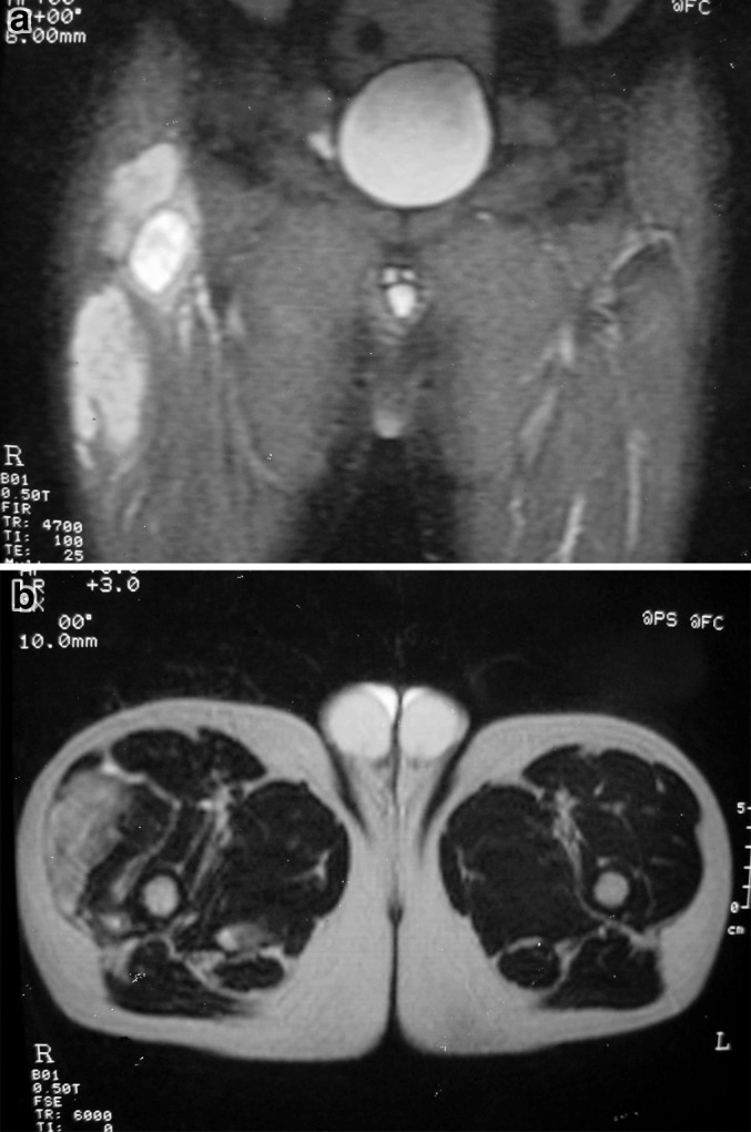
Fig. 5.

aCoronal T-2 weighted magnetic resonance image of the pelvis showing a soft tissue mass at the right hip and proximal thigh, associated with destruction and significant edema of the right trochanter. b Axial T-2 weighted magnetic resonance image showing a soft tissue mass of the right proximal thigh
