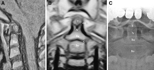
Fig. 1.
a Sagittal T1-weighted magnetic resonance imaging (MRI) in a 3 years old pediatric individual shows the tip, neck, and base of the odontoid process (T tip of the odontoid, N the neck of the odontoid, Ba the base of the odontoid, Bo the body of C2, one arrow shows dentocentral synchondrosis, double arrow shows apicodental synchondrosis). b. Coronal T1-weighted MRI of a pediatric case shows the segments of the odontoid process (T tip of the odontoid, N the neck of the odontoid, Ba the base of the odontoid, Bo the body of C2, one arrow shows dentocentral synchondrosis, double arrow shows apicodental synchondrosis). c Segmentation of an adult odontoid process in a direct X-ray (T tip of the odontoid, N the neck of the odontoid, Ba the base of the odontoid, Bo the body of C2)

