
Fig. 1.
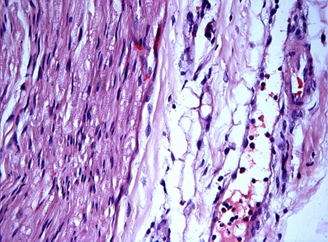
Control group. Control sciatic nerve. Nerve fascicle (left) and perineurium (right). Note the presence of some neutrophylic granulocytes in the interstitium near a postcapillary venule and adhesion of neutrophylic granulocytes to the endothelium of the venule. H&E staining. Magnification 400×