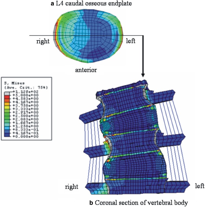
Fig. 6.
Stress distributions in right lateral bending at 10 N m with precompression loading in pediatric models. a Top view of the L4 caudal osseous endplate, b anterior view of the coronal section of the model through the middle of vertebral body as indicated in a. In a, both right and left lateral corner are shown to be highly stressed due to compression and traction force, respectively. The stress distribution pattern in b indicates the high loading at the osseous endplate and apophyseal bony ring

