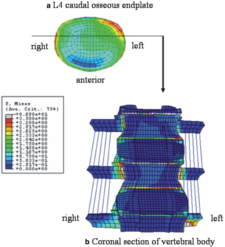
Fig. 7.
Stress distributions in right axial rotation at 10 N m with precompression loading in pediatric models. a Top view of the L4 caudal osseous endplate, b anterior view of the coronal section of the model through the anterior one-third of vertebral body as indicated in a. In a, left posterior–lateral corner is shown to be highly stressed. The stress distribution pattern in b indicates the high loading at the osseous endplate and apophyseal bony ring

