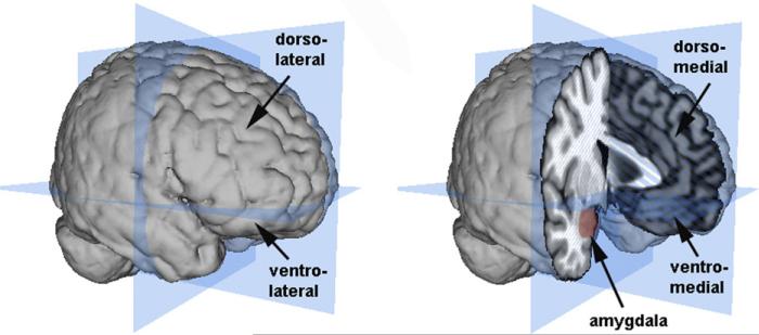
Fig. 1.
Illustration of the amygdala and the major divisions of the PFC. The planes (in blue) show the major dorsal/ventral and anterior/posterior divisions of the brain. The lateral PFC is shown on the left and the medial PFC and amygdala are shown on the right. Brain images and surface constructions were created using Mango (Research Imaging Center, UTHSCSA; http://ric.uthscsa.edu/mango/mango.html) and a Montreal Neurological Institute standard brain.

