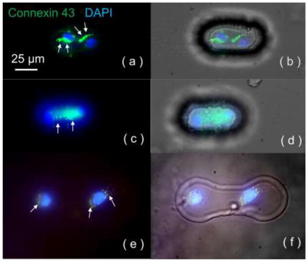
Figure 4.
Connexin 43 staining for contact-promotive/preventive biochips. The DAPI staining (blue) on the nucleus indicates two cells in one microwell, and Cy3 staining (green) indicates connexin 43 expression. (a, b) Junctional connexin 43 distribution at the cell-contact area was found in contact-promotive biochips; (c, d) Diffusive connexin 43 was expressed throughout the cell bodies in contact-promotive biochips. (e, f) In the contact-preventive biochips, portions of surviving heterotypic cell pairs were found to have diffusive connexin 43 expression. White arrows indicated the location of connexin 43.

