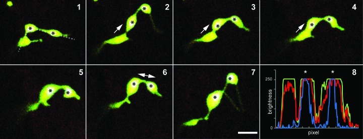
Figure 2. Non-green plastids like etioplasts and leucoplasts lack large rigid internal structures and this allows their shape to be more flexible than mesophyll chloroplasts. The morphological flexibility and the formation of a constricted isthmus in late stages of plastid division frequently gives the impression of two or more plastids being connected by stromules. Because most of these plastids are missing grana organization, the identification of the main plastid body in pleomorphic tubules is difficult. Seven frames (1–7) from a 5 min time series depicting a plastid in a mature A. thaliana root epidermal cell show plastid pleomorphy. At the beginning (1), three bulbous regions are connected by tubules, suggesting plastids interconnected by stromules. However the chlorophyll signal, which can be detected in plastids in mature roots of in vitro, light grown plants, is observed in only two of the plastid shaped bulbous regions (marked by '*'). The RGB-intensity plot (see 8; blue line denotes chlorophyll fluorescence) confirms the observation of two plastids only in the group of bulbous dilations. During the time lapse series the dilation without chlorophyll signal fused with one of the chlorophyll signal-containing bulbs (2–4), indicating that both bulbs were indeed parts of the same plastid. (8) The RGB-intensity plots along the dotted line in (1) illustrate the even distribution of photoconverted red and non-photoconverted green mEosFP (visible in the merged images 1–7 as yellowish color) and clearly proves the presence or absence of chlorophyll in the bulbs (overlay of blue chlorophyll signals with green and red result in white sectors ‘*’). Prominent movements of bulbs are indicated by arrows. Size bar = 5µm.

An official website of the United States government
Here's how you know
Official websites use .gov
A
.gov website belongs to an official
government organization in the United States.
Secure .gov websites use HTTPS
A lock (
) or https:// means you've safely
connected to the .gov website. Share sensitive
information only on official, secure websites.
