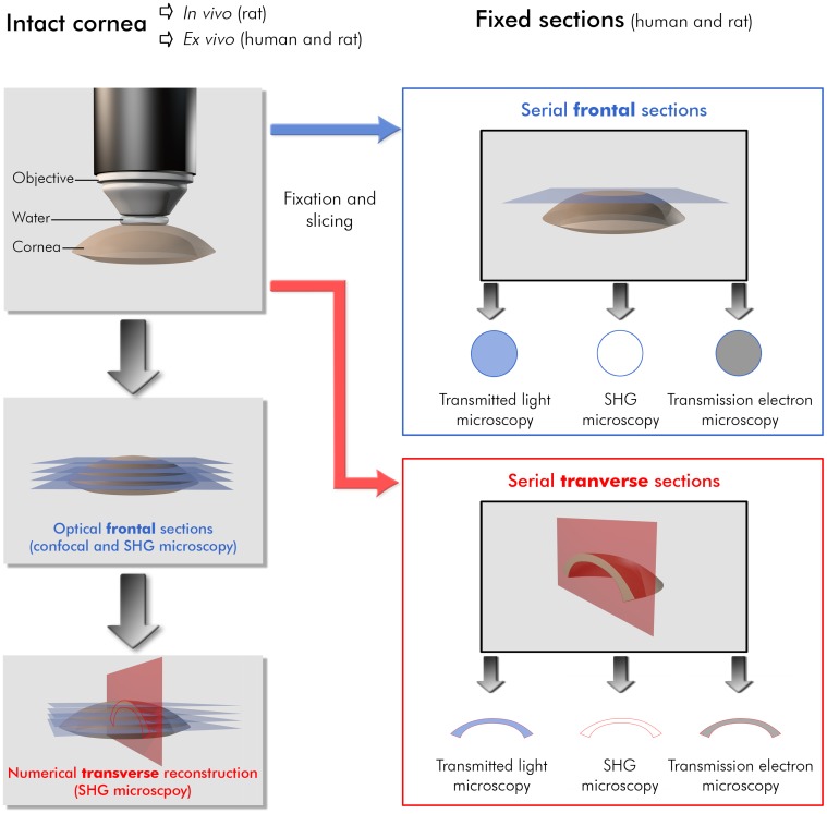
Figure 1. Methodology to compare the different imaging techniques on rat and human corneas.
Cornea preparation, orientation of the different histological and numerical sections and imaging geometry are indicated for each imaging technique. Histological sections are unstained for SHG microscopy, stained with toluidine blue for transmitted light microscopy and stained with uranyl and lead citrate solutions for transmission electron microscopy.

