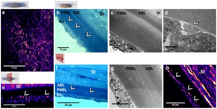
Figure 4. Multimodal imaging of the DM from the same diabetic human cornea.
(a–d) Frontal and (e–h) transverse sections. (a, e) SHG microscopy of intact cornea: (a) frontal optical section, (e) transverse numerical reconstruction. (b, f) Transmitted light microcopy of stained histological sections, where the abnormalities are visible with few contrast. (c, d, g) TEM views of (c, g) the entire DM and (d) its posterior part and the endothelium: long-spacing collagen appears to be synthesized by the endothelial cell. (h) SHG imaging of the same transverse histological section, where fibrillar collagen is clearly identified in the DM. St: stroma, ABL: anterior banded layer, PNBL: posterior nonbanded layer, En: endothelium. White arrows indicate collagen abnormal deposits in the DM.

