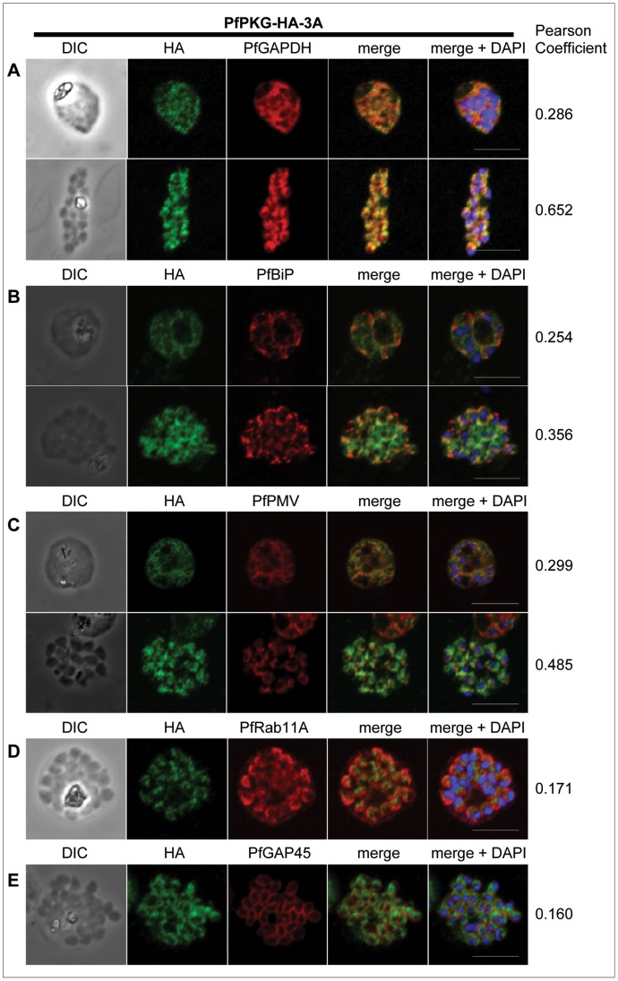
Figure 2. Subcellular location of PfPKG in mature schizonts.
Dual immunofluorescent detection of PfPKG-HA in fixed smears of early and late schizonts of the PfPKG-HA-3A clone together with (A) PfGAPDH [30], (B) PfBiP [32], (C) PfPMV [31], (D) PfRab11A [33] and (E) PfGAP45 [34]. Representative images are shown for each antibody, together with bright field images (first column) and parasite nuclei stained with DAPI (in the merged image). Bars ∼5 µM. To quantify co-localisation, Pearson coefficients [36] of the individual stains were calculated using Imaris image analysis software (Bitplane).

