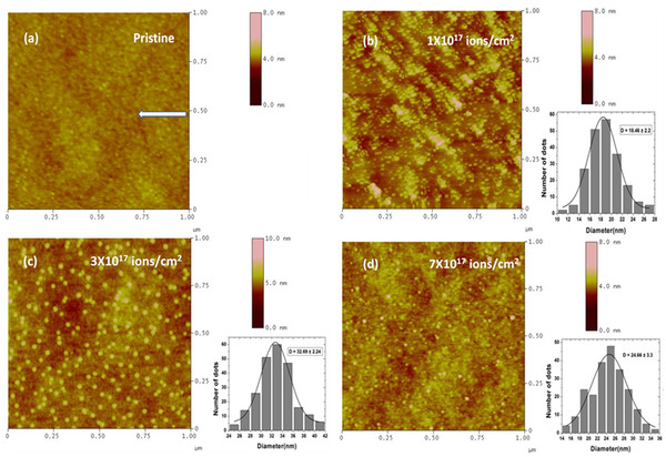
Figure 5.

AFM micrographs. (a) Pristine; 50-keV Ar+-irradiated substrates of GaAs (100) at an angle of 50° with respect to surface normal at different fluences: (b) 1 × 10¹⁷, (c) 3 × 10¹⁷, and (d) 7 × 10¹⁷ ions/cm2. The arrow in the figure indicates the projection of ion beam direction on the surface. Nano-dot size distribution is shown in the insets.
