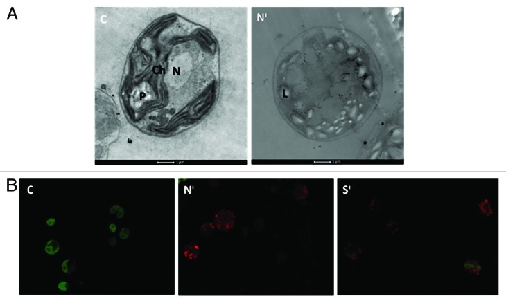
Figure 1. Transmission electron microscopy (A) and Confocal fluorescence microscopy (B) images of control, N-starved and S-starved C. reinhardtii cells sampled on fifth day of incubation. In the fluorescence images green represents chlorophyll autofluorescence and light red represents Nile Red fluorescence. Abbreviations: C, Control; Nᶦ, N-deprived cells; Sᶦ, S-deprived cells; Ch, Chloroplast; P, Pyrenoid; N, Nucleus; L, lipid bodies.

An official website of the United States government
Here's how you know
Official websites use .gov
A
.gov website belongs to an official
government organization in the United States.
Secure .gov websites use HTTPS
A lock (
) or https:// means you've safely
connected to the .gov website. Share sensitive
information only on official, secure websites.
