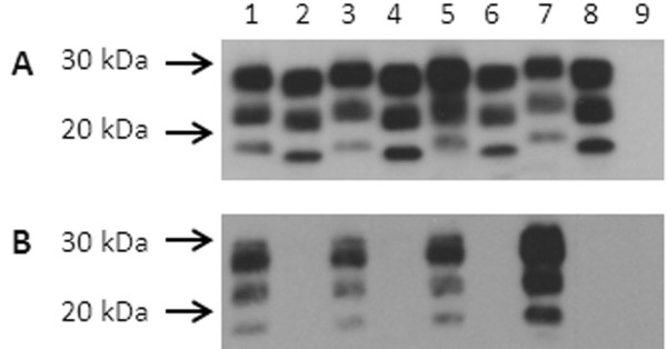
Figure 4 .
Co-migration of brain homogenates from Tg338 mice inoculated by the intracerebral route or the intraperitoneal route. Brain homogenate from sheep 3178 (lanes 1, 3, 5 and 7, 350 μg starting wet weight per lane) and Tg338 mice (100 μg starting wet weight per lane) inoculated by the intracerebral route and assayed at passage 2 (lane 2) or passage 1 (lane 4). The same homogenate was inoculated intraperitoneally in Tg338 mice; first passage brain (lane 6) and spleen (lane 8) homogenates were assayed. Lane 9, brain homogenate (200 μg starting wet weight) from uninoculated Tg338 mouse. All tissues were digested with proteinase K (100 μg /ml final concentration for murine tissues and 50 μg/ml final concentration for ovine tissues). Filter was probed with mAb F99/97.6.1. Molecular mass markers are shown on the left.

