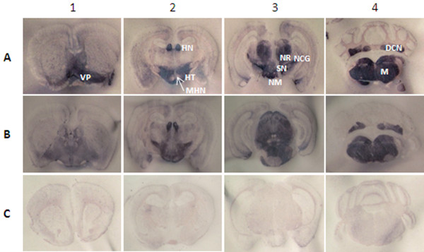
Figure 7 .
Distribution of PK-resistant PrP labeling of PET blots. Shown is the neuroanatomic distribution of PrPSc at 4 different brain levels of P2 Tg338 mice representing inoculum groups 30–75 (A) and 3178 (B), and an age-matched uninoculated control group mouse (C). Brain sections were dissected at the levels of frontal cortex (level 1), hypothalamus (level 2), hippocampus (level 3) and cerebellum-medulla (level 4). Blots were probed with mAb F99/97.6.1. VP, ventral pallidum; HT, hypothalamus; MHN, medial hypothalamic nucleus; HN, medial habenular nucleus; NM, nucleus mammillaris; NR, nucleus rubor; SN, substantia nigra; NCG, nucleus corporis geniculati; DCN, deep cerebellar nuclei; M, medulla.

