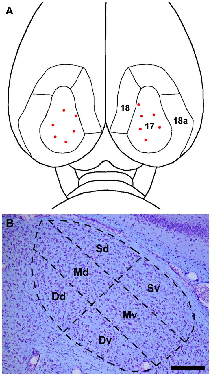
Figure 1. Dorsal view of the rat brain showing visual cortical areas 17, 18, and 18a.
(A) The sites of biotinylated dextran amine (BDA) injections are represented by red dots. (B) A schematic drawing representing a coronal section of the dLGN near the rostro-caudal midpoint of the nucleus, A–P plane 5, superimposed on a corresponding cresyl violet stained section of the dLGN. The dLGN is divided arbitrarily into six sectors: Sd, superficial dorsal; Sv, superficial ventral; Md, mid-dorsal; Mv, mid-ventral; Dd, deep dorsal; Dv, deep ventral. Scale bar: 3.0 mm for A and 250 µm for B.

