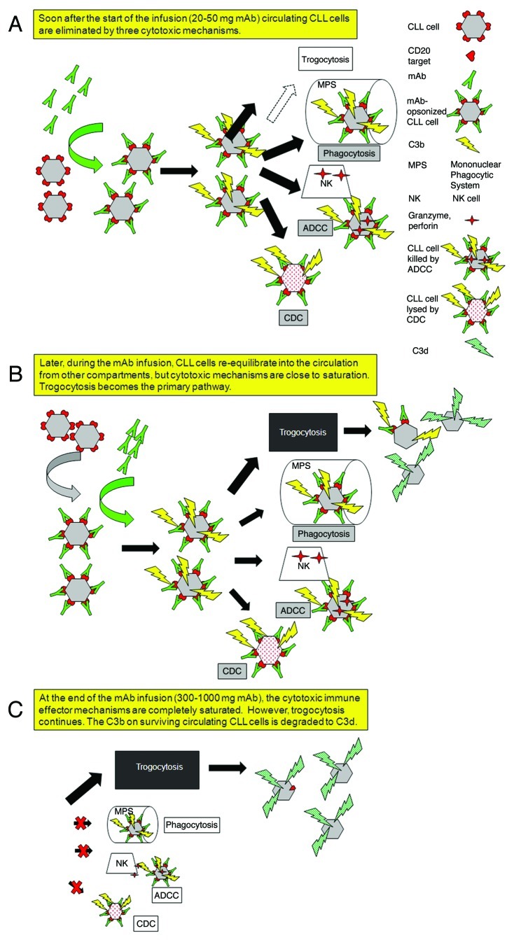
Figure 1. Schematic illustration of the sequence of events that occurs when CLL patients receive intravenous infusions of large quantities of Type I CD20 mAbs. (A) Several of the body’s immune effector mechanisms promote a high level of clearance and destruction of circulating CLL cells after infusion of the first 20–50 mg of the Type I CD20 mAb. (B) Later, after a first wave of clearance, a substantial number of CD20+ CLL cells have re-equilibrated into the bloodstream from other compartments. The cells are opsonized by mAb, but the cytotoxic mechanisms are less effective, and an alternative reaction predominates: trogocytosis (shaving) of bound mAb and CD20 by fixed cells that express Fcγ receptors. (C) After the infusion is complete, the effector mechanisms are nearly exhausted or saturated, but trogocytosis continues. Although the complement titer is substantially reduced, there is sufficient residual complement activity that the cells are covalently opsonized with C3 activation fragments (which decay to C3d) before they lose CD20. These C3d-opsonized “low CD20” CLL cells are not cleared, and can remain in the circulation for weeks to more than one month.

An official website of the United States government
Here's how you know
Official websites use .gov
A
.gov website belongs to an official
government organization in the United States.
Secure .gov websites use HTTPS
A lock (
) or https:// means you've safely
connected to the .gov website. Share sensitive
information only on official, secure websites.
