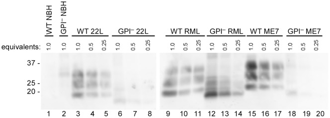
Figure 3. PrPRes levels in brains of GPI− and WT mice infected with multiple mouse-adapted scrapie strains.
Normal brain homogenate as well as 22L, RML and ME7-infected brain homogenates were compared by immunoblotting. The sample brain equivalents were loaded into each lane. Lanes 1–2: WT and GPI− NBH undiluted, respectively; Lanes 3–8: WT and GPI− 22L BH undiluted and serially diluted 2-fold and 4-fold; Lanes 9–14: WT and GPI− RML BH undiluted and serially diluted 2-fold and 4-fold; Lanes 15–20: WT and GPI− ME7 BH undiluted and serially diluted 2-fold and 4-fold. A final concentration of 20 µg/mL PK was used to digest brain homogenates. Bands were detected with monoclonal antibody 6D11 as described in materials and methods.

