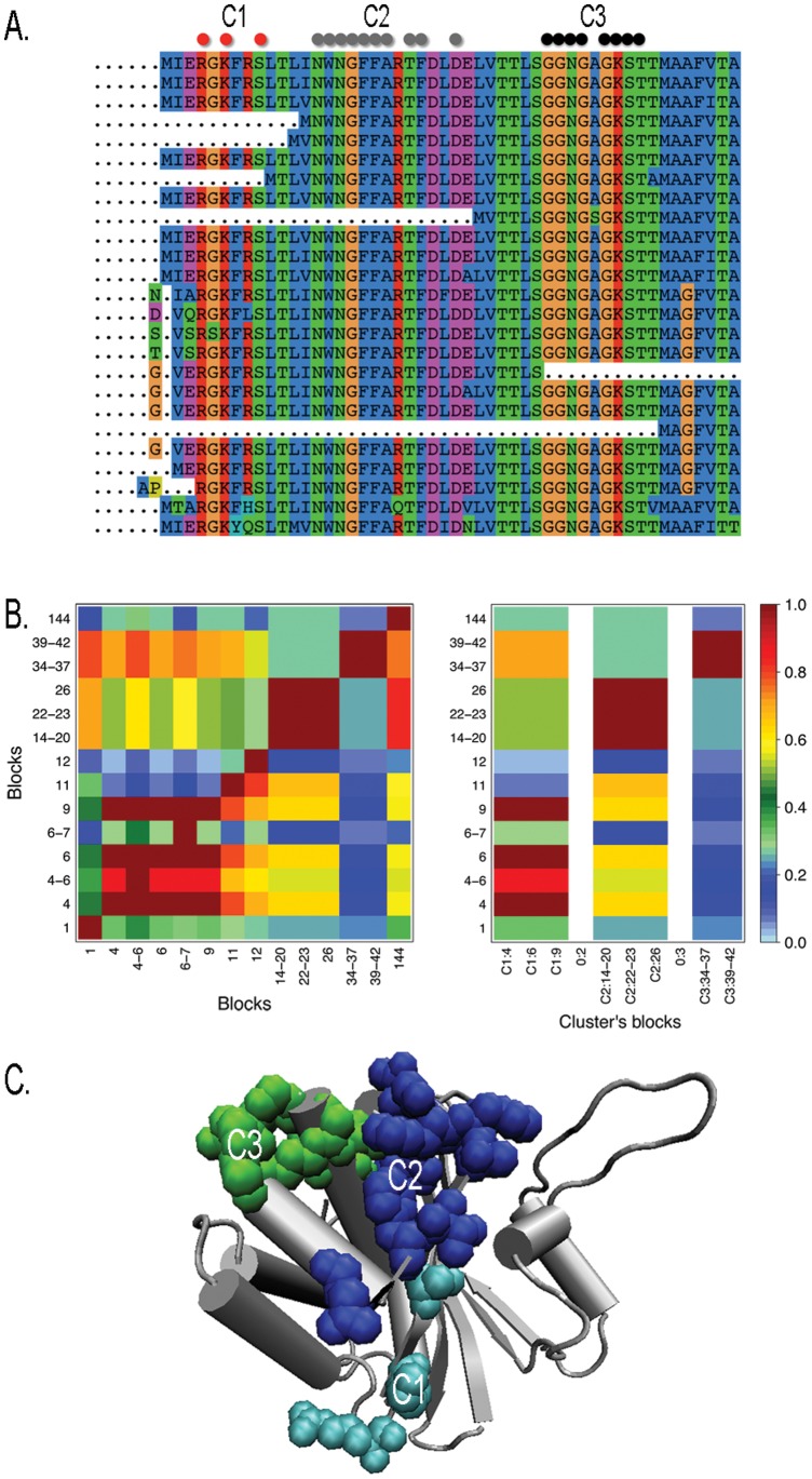
Figure 2. The Walker-A motif in MukB proteins.
Display of a subset of the full sequence alignment (made of 200 members, with 84% sequence identity) used to analyze coevolution in the MukB family. Sequences are truncated on the right. Three clusters are indicated by colored dots (top). B. Matrix of coevolution scores between blocks of dimension 0 (left) and clustered coevolution score matrix highlighting 3 resulting clusters (right). See Text S15. C. Clusters C1–C3 are plot in the structure (1qhl:A). Walker-A (C3) is colored green.

