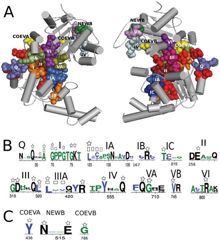
Figure 5. AATPase protein Upf1.
A: Upf1 protein structure (2gjk) where all known motifs of the family appear in distinguished colors together with their extensions (in red), one new motif (green, named NEWB) and two more coevolving residues (yellow, named COEVA and COEVB), not know to play a functional role in Upf1, have been identified by BIS. Two domains (left) and their backside (right) are shown (Texts S8 and S15). B: protein logos of all known motifs and their extensions. Residues belonging to the extensions are marked by a square and a star marks those that are coevolving. BIS analysis detects 26 coevolved positions (20 for  and 6 for
  ) over 677 alignment positions. C: protein logos of the new motif and the two coevolved positions.

