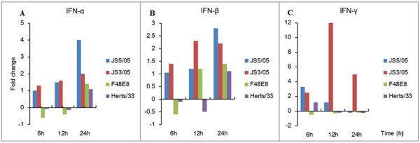
Figure 3.
Determination of mRNA expression of IFN-α, IFN-β and IFN-γ genes in the splenocytes. Profiles of IFN-α, IFN-β and IFN-γ gene expression were shown in panel A, B and C respectively. The standard curve method was used to analyze the fold change of relative gene expression levels between mock-infected and infected cells. Relative expression levels were normalized to the internal β-actin. The data are the mean fold change ± standard deviation (SD).

