
Figure 1.
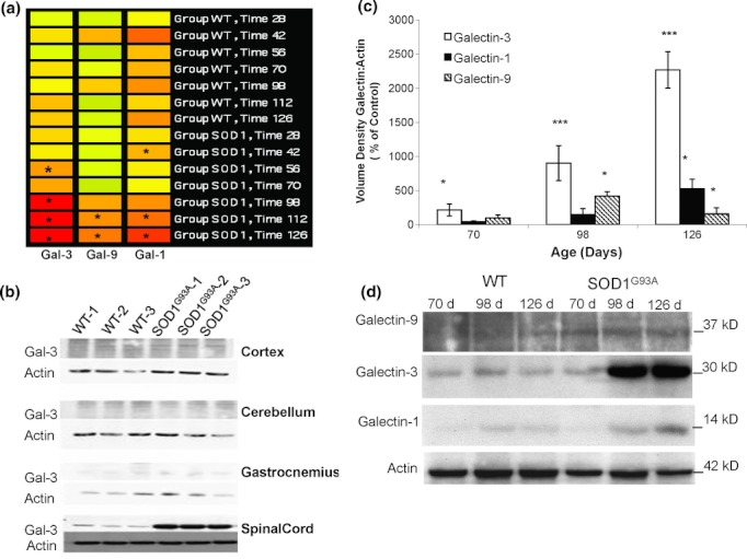
Galectins are specifically and differentially expressed in the spinal cord of mice with chronic motor neuron disease. (a) Heat map of galectin mRNA transcripts expressed over time as detected by microarray analyses of spinal cords from B6SJL SOD1 G93A versus control B6SJL (WT) mice (n = 3 per genotype). Each column represents the signal from a specific galectin probeset (1426808_at=galectin-3, 1421217_a_at=galectin-9, 1419573_a_at=galectin-1), and each line of bars represents mouse age (in days). Expression signal intensities are depicted as colors ranging from high (orange/warm) to low (green/cool), relative to the signal of all controls combined. Asterisks indicate significant differences from controls of equivalent age (P < 0.05 by Student's t-test comparison). In this strain, disease onset occurs at ∼98 days of age, and end-stage disease at ∼130 days of age. (b) Elevated levels of galectin-3 are restricted to the spinal cord, rather than other CNS regions or muscle. Galectin-3 protein levels were assessed with Western blots of homogenates prepared from the cortex, cerebellum, gastrocnemius muscle, and spinal cord of 126-day-old wild-type (WT) and B6SJL SOD1 G93A mice (n = 3 per genotype). (c) Expression of galectin-3 is elevated early in the spinal cord during motor neuron disease progression, followed by increases in galectins-9 and -1. Western blots were prepared from spinal cord homogenates of 70-, 98-, and 126-day-old B6SJL SOD1 G93A mice and age-matched strain controls (n = 3 per genotype). Blots were quantified, and optical densities of galectins expressed as the galectin:actin ratio as a percent of age-matched strain controls (*P < 0.05; ***P < 0.001 compared with controls by Student's t-test). Representative blots are shown below (d).