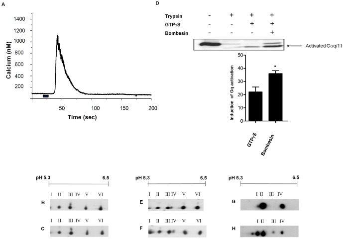
Figure 4. Sodium vanadate treatment mimics PMT effects on Gαq and Gαi.
(A) Indo-1 AM labelled Swiss 3T3 cells were treated with 30 nM bombesin for 10 s (marked with solid bar beneath trace) and intracellular Ca2+ release was measured. Membrane proteins from Swiss 3T3 cells that were either (B) untreated or (C) treated with 30 nM bombesin for 1 min were separated by 2-D gel electrophoresis and Western blotted with anti-Gαq/11 antibody. (D) Swiss 3T3 membrane proteins were incubated in the presence or absence of 30 nM bombesin for 20 min with or without 0.05 nM GTPγS. The proteins were then analysed for trypsin protection as described under Materials and Methods, and activated Gαq/11 was separated by SDS PAGE and Western blotted with anti-Gαq/11 antibody. Quantification of activated Gαq/11 (lower panel) was determined by densitometric scanning and these data were analysed using factorial analysis of variance (ANOVA). The induction of activation shown is relative to the density of the band without GTPγS or bombesin. Bombesin significantly enhanced GTPγS binding to Gαq/11 (* p = 0.002). Membrane proteins from Swiss 3T3 cells were incubated with (E) 1 mM sodium vanadate for 20 min at 37°C or (F) 1 mM sodium vanadate and 30 nM bombesin for 20 min at 37°C, proteins were separated by 2-D gel electrophoresis and Western blotted with anti-Gαq/11 antibody. Membrane proteins from Swiss 3T3 cells were incubated (G) without or (H) with 1 mM sodium vanadate for 20 min at 37°C, proteins were separated by 2-D gel electrophoresis and Western blotted with anti-Gαi-1-3 antibody. Samples from at least 3 independent membrane preparations were resolved with similar results.

