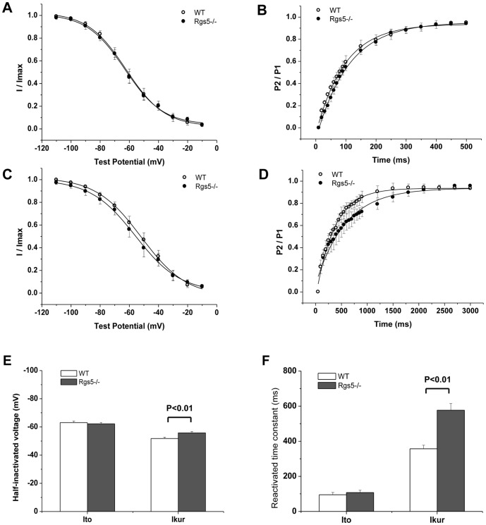
Figure 7. Channel kinetics of Ito and IKur were analyzed in Rgs5−/− and WT cardiomyocytes, respectively.
The steady-state inactivation (A) and reactivation (B) of curves in Ito plotted similar tendency between Rgs5−/− and wild-type atrial myocytes. However, the property of IKur in Rgs5−/− cardiomyocytes showed markedly modification with significant difference of half-inactivated voltage (V1/2) (C) and reactivated time constant (τ) (D). Histogram summarized the statistical significance of V1/2 and τ in WT and Rgs5−/− groups (E, F).

