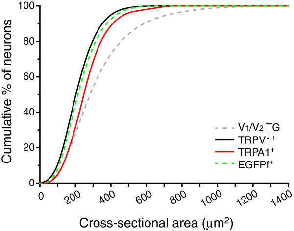
Figure 8.
Overlay of the cumulative distributions of the TRPV1+, TRPA1+and TRPM8-expressing neurons in the V1/V2divisions of the TG. Cumulative distribution of the cross-sectional areas of the total TG neurons in the V1/V2 divisions (dashed gray line, the same neurons as in Figure 1F), the TRPV1+ neurons (solid black line, the same neurons as in Figure 5E), the TRPA1+ neurons (red line, the same neurons as in Figure 6E), and the TRPM8-expressing neurons (dashed green line, the same neurons as in Figure 7E). A Kruskal-Wallis ANOVA with Dunn’s post hoc test reveals that the sizes of TRPV1+ neurons are the smallest of the three populations of TG neurons (p < 0.001 and p < 0.05 compared with the TRPA1+ and EGFPf+ groups, respectively). In contrast, the sizes of TRPA1+ neurons are significantly larger than those of the TRPV1+ and TRPM8-expressing neurons (p < 0.001 and p < 0.05, respectively).

