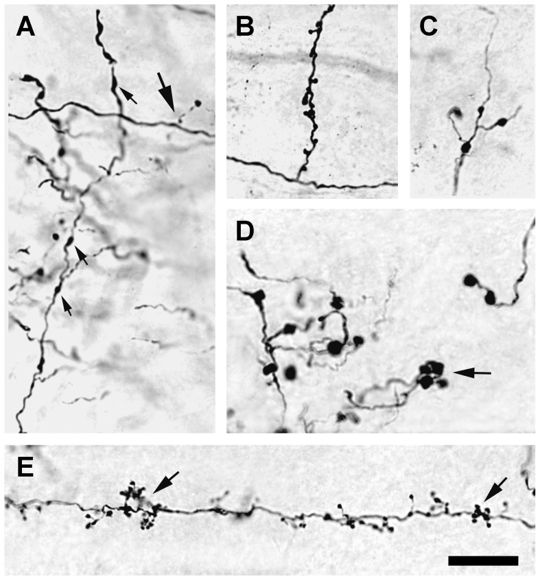
Figure 7. Micrographs of axons 3 days after a 0.1 µL injection of 3,000 MW BDA.
(A) Corticocortical axons with varicosities (upward arrows). Occasionally, short branches that terminate in round spherules (large arrow) also were seen. (B) Corticogeniculate axon with an atypically long side branch studded with boutons. (C) Corticothalamic axon in the lateral dorsal nucleus, with small diameter varicosities (2–3 µm) and short appendages. (D) Corticothalamic axons in the lateral posterior nucleus of the thalamus with many large varicosities (>4 µm in diameter) and boutons that often were seen in clusters (arrow). (E) Typical fine caliber, corticogeniculate axon is beaded with small varicosities, and supports many dense, short appendages that end in boutons (arrows) that frequently are arrayed in clusters. Scale bar: 20 µm.

