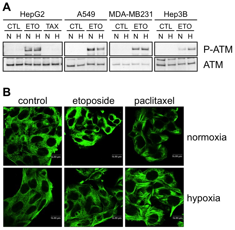
Figure 2. Effects of hypoxia on drug-induced damage.

(A) Effect of hypoxia, etoposide and paclitaxel on the abundance and phosphorylation of ATM. HepG2, A549, MDA-MB231 and Hep3B cells were incubated 1 hour under normoxia (N, 21% O2) or hypoxia (H, 1% O2) in the presence or not (CTL) of etoposide (ETO, 100 µM in Hep3B cells and 50 µM in the other cell types) or paclitaxel (TAX, 10 µM) in HepG2 cells. ATM and P-ATM were detected in nuclear protein extracts by western blotting using specific antibodies. One experiment representative out of three. Uncropped western blots are presented in Figure S1. (B) Effect of hypoxia, etoposide and paclitaxel on microtubules. HepG2 cells were incubated 16 hours under normoxia (21% O2) or hypoxia (1% O2) in the presence or not of etoposide (50 µM) or paclitaxel (10 µM). After the incubation, cells were fixed, permeabilised and stained for alpha-tubulin using a specific antibody. Observation was performed using a confocal microscope with a constant photomultiplier.
