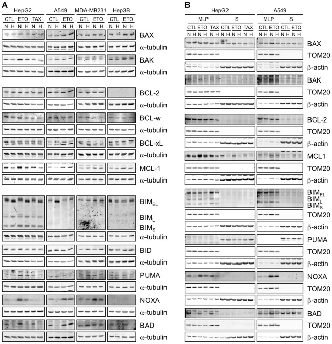
Figure 6. Effect of hypoxia, etoposide and paclitaxel on the abundance and localisation of BCL-2 family proteins.
HepG2, A549, MDA-MB231 and Hep3B cells were incubated 16 hours under normoxia (N, 21% O2) or hypoxia (H, 1% O2) in the presence or not (CTL) of etoposide (ETO, 100 µM in Hep3B cells and 50 µM in the other cell types) or paclitaxel (TAX, 10 µM) in HepG2 cells. (A) Proteins were detected in total cell extracts by western blotting, using specific antibodies. alpha-tubulin was used as loading control. One experiment representative out of three. Uncropped western blots are presented in Figure S1. (B) After the incubation, subcellular fractionation was performed and proteins were detected in the MLP (mitochondria-lysosome-peroxisome) and S (cytosolic) fractions by western blotting, using specific antibodies. TOM20 and β-actin were used as loading controls for the MLP and S fractions respectively. One experiment representative out of three. Uncropped western blots are presented in Figure S1.

