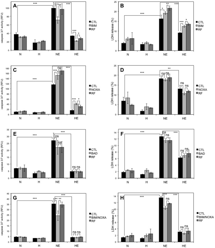
Figure 7. Effect of BH3-only proteins silencing on the etoposide-induced cell death.
. HepG2 cells were transfected with 50 nM BIM (A, B), NOXA (C, D) or BAD (E, F) siRNAs, or 25 nM BIM combined with 25 nM NOXA (G, H), or 50 nM RISC-free (RF) control siRNA or left untransfected (CTL) for 24 hours. 6 hours later (or 30 hours later for A and B), cells were incubated under normoxia (N, 21% O2) or hypoxia (H, 1% O2) with (ETO) or without (CTL) etoposide (50 µM) for 16 (A, C, E, G) or 40 hours (B, D, F, H). (A, C, E, G) Caspase-3/7 activity was assayed by measuring the fluorescence of free AFC released from the cleavage of the caspase-3/7 specific substrate Ac-DEVD-AFC. Results are expressed in relative fluorescence units (RFU) as means ±1 SD (n = 3). (B, D, F, H) LDH release was assessed. Results are presented in percentages as means ±1 SD (n = 4, but n = 3 in B for the condition HE CTL and in f for the condition H BAD). (A-H) Statistical analysis was carried out with ANOVA 1. ns: non-significant; *: P<0.05; **: P<0.01; ***: P<0.001.

