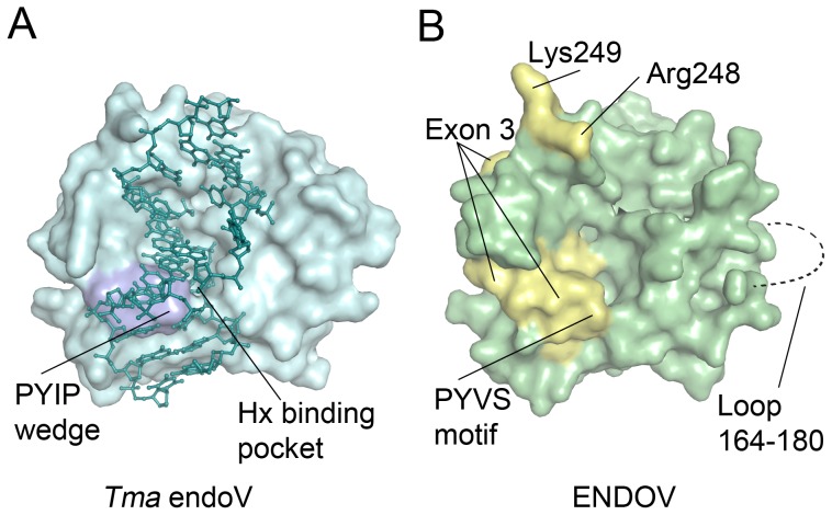
Figure 7. Location of structural elements in endonuclease V.
(A) Structure of Tma endoV binding to deaminated DNA (PDB code 2W35 [23]) showing the central location of the strand-separating wedge (PYIP motif) close to the damage recognition pocket. (B). Homology model of human ENDOV showing the location of mutated residues Arg248 and Lys249 (yellow), as well as residues forming the PYVS motif in exon 3 (yellow), which were also mutated. A loop comprising residues 164–180 could not be reliably modelled and is not included (dashed line).

