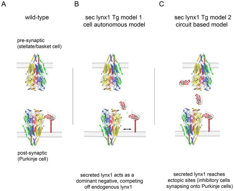
Figure 4. Two schemes for possible modes of action of secreted lynx on nicotinic receptors within synapses.
(A) Schematic diagram of a synapse in WT mice. Schematic model of nicotinic receptors and lynx1 interaction at a Purkinje cell synapse. Models are based on the crystal structure of AChBP [57] and the NMR structure of lynx1 [56]. Lynx1 is depicted as binding at the subunit interface of the pentameric channel, based on α-bungarotoxin binding. Lynx1 expression is expressed in the post-synaptic cell (Purkinje cell), and is not expressed in the presynaptic neuron (stellate/basket neuron). Tethered to the membrane by it GPI-anchor, lynx is depicted as having access to a nicotinic receptor binding site at the post-synaptic face only. (B) Schematic model 1 of sec-lynx1 function – cell autonomous, dominant negative model. Schematic representation of Purkinje cell synapses in L7-sec-lynx1 Tg mice. Binding of secreted lynx1 to the same subunit interface of the nicotinic receptor could compete off the binding of native full-length GPI-anchored lynx1, and thereby exert a dominant negative effect. This model implies that the secreted version of lynx1 has either no effect or a differential function as compared to the membrane bound version of lynx1, but maintains nicotinic receptor binding capability. (C) Schematic model 2 of sec-lynx1 function – circuit based, ectopic expression model. In this model, the soluble lynx1 secreted from the post-synaptic Purkinje cells diffuses extracellularly, accessing nicotinic receptors located on terminals of pre-synaptic neurons (stellate/basket cell). In this model, the ectopic expression of lynx1 in pre-synaptic sites can lead to suppression of activity or neurotransmitter release, leading to dis-inhibition onto Purkinje cells. This dis-inhibition can lead to alterations in excitatory/inhibitory balance and motor learning.

