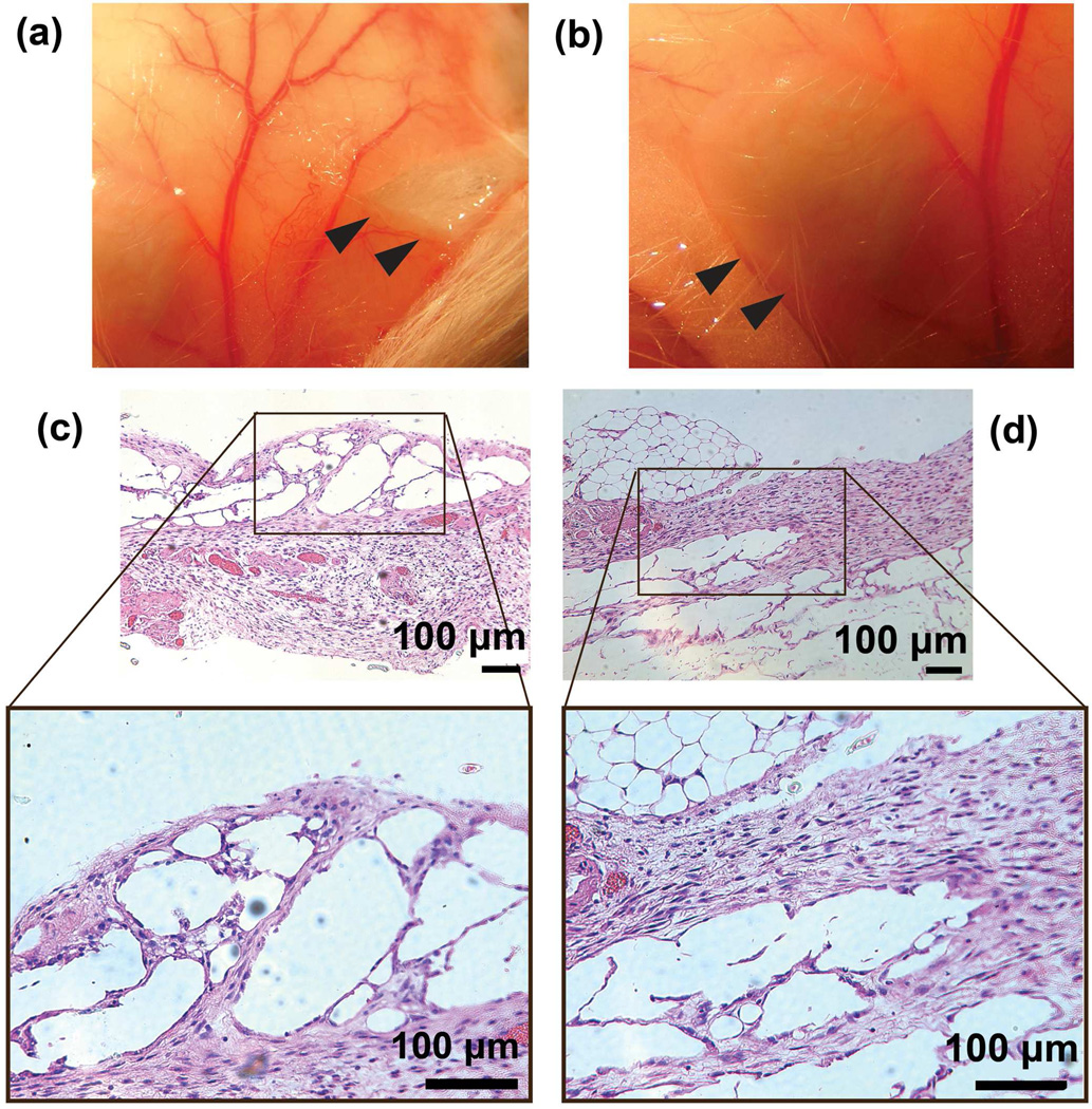
Figure 4.
(a) and (b) are the photographs of in situ formed conductive hydrogels containing 1 wt% of PPy NPs after a subcutaneous injection in FVB mice. The hydrogel formed subcutaneously in the mouse and showed a spherical to ovoid shape after 1 week (a) and 2 weeks (b) healing. The gel was removed from the mouse on week 1 and 2. Black arrows indicated the implants. (c) and (d) are H&E stained images of the conductive hydrogels after subcutaneous implantation in an FVB mouse at 1 week (c) and 2 weeks (d). H&E stain cells could be observed in the hydrogel area.

