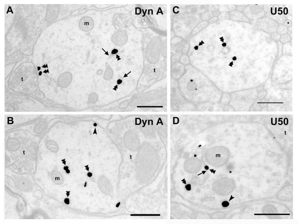
Fig. 3. Subcellular distribution of KOPR-IR after acute agonist treatment.
Sections from rats that received an intrathecal injection of dynorphin A (1-17) (15 nmole) (A, B) or U50,488H (100 nmole) (C, D). The arrows point to the endosomes with KOPR. Double arrowheads point to immunogold-silver labeling in the cytoplasm, while single arrowheads point to immunogold-silver labeling on the plasma membrane. t, axon terminal; m, mitochondria. Scale bar = 0.5 μm.

