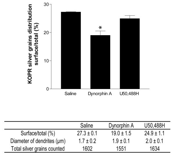
Fig. 4. Quantification of association of KOPR-IR with the plasma membrane in dorsal horn dendrites of rat lumbar spinal cords.
The diameters of the sampled dendrites and the number of total silver grains in each group were shown. Bars represented the mean percentage of KOPR-silver grains associated with the plasma membrane in rats intrathecally injected with dynorphin A (15 nmole), U50,488H (100 nmole) or saline. Values are mean ± SEM of data obtained from 3 rats per group, at least 150 dendrite profiles per rat.*, p<0.05, compared with saline group by Fisher’s exact test using the silver grain numbers categorized into on the surface or in the cytoplasm.

