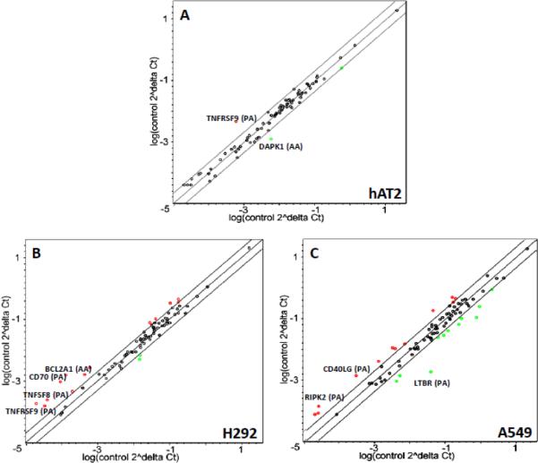
Figure 8.
Scatter plot analysis of the contribution of HSulf-1 to the apoptotic effects of cadmium. (A) hAT2, (B) H292, and (C) A549 cells were exposed to cadmium for 48 hours after adenovirally-transduced over-expression of lacZ or HSulf-1. Genes significantly up- or down-regulated (more than 2-fold) by cadmium in HSulf-1-expressing cells compared to those regulated by cadmium in lacZ-expressing cells are shown in red or green, respectively, and the most significant genes are labeled: (PA) pro-apoptosis; (AA) anti-apoptosis.

