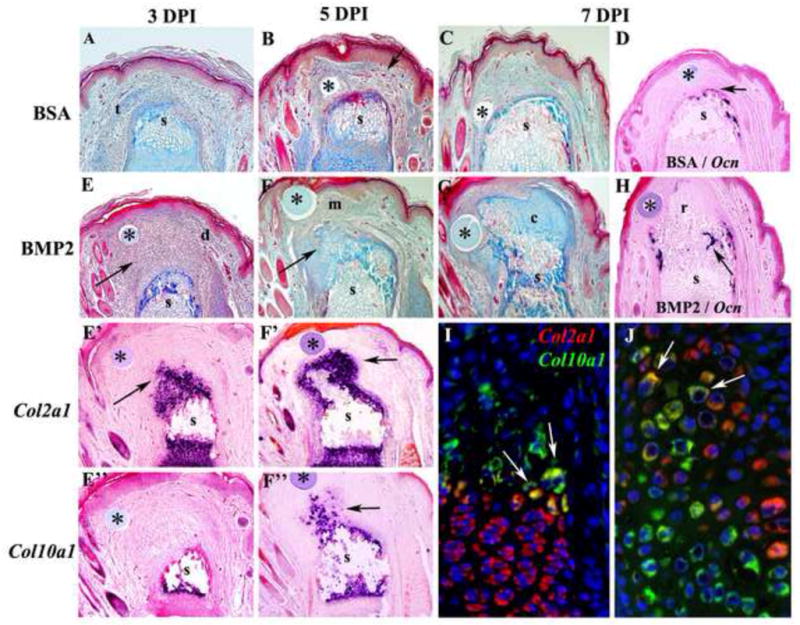
Figure 2. Endochondral ossification during BMP2 induced regeneration.

Distal is toward the top in all images and microcarrrier beads are indicated by *. A–C) Histological analysis of control BSA-treated P2 amputations. A) At 3 DPI there is evidence of wound contraction based on differentiated epidermal structures along the periphery of the stump (s) and encroachment of a severed tendon (t) over the amputated skeletal stump. B) At 5 DPI the control amputation wound associated with a BSA bead is composed of vascularized (arrow) fibrous connective tissue covering the skeletal stump (s). C) At 7 DPI the amputation wound associated with the control BSA bead continues to accumulate fibrous connective tissue that covers the skeletal stump (s). D) Section in situ hybridization at 7 DPI showing cells expressing the osteoblast marker Osteocalcin forming a cap over the amputated skeletal stump (s). E–G) BMP2-treated P2 amputations. E) Histological section at 3 DPI showing the accumulation of mesenchymal cells (arrow) distal to the amputated skeletal stump (s) and associated with the BMP2 bead. The mesenchymal cell mass is flanked with connective tissue of the dermis (d) and is oriented in the direction of the BMP2 bead. E′) Section in situ hybridization at 3 DPI showing Col2a1 transcripts (arrowhead) associated with cells just distal to the skeletal stump (s) and not associated with the BMP2 bead. E″) Section in situ hybridization for Col10a1 transcripts showing the absence of hypertrophic chondrocytes distal to the amputated stump (s) at 3 DPI. F) Histological section at 5 DPI showing a reduced population of mesenchymal cells (m) associated with the BMP2 bead, and chondrocytic tissues (arrow) contiguous with, and distal to, the amputated skeletal stump (s). F′) Section in situ hybridization for Col2a1 transcripts (arrow) showing that chondrocytes associated with the skeletal stump (s) extend distally toward the BMP2 bead. F″) Section in situ hybridization for Col10a1 transcripts (arrow) showing that hypertrophic chondrocytes distal to the amputated stump (s) are present at 5 DPI. G) Histological section at 7 DPI showing few distal mesenchymal cells, and a large mass of chondrocytic tissue (c) contiguous with, and distal to, the amputated skeletal stump (s) that is associated with the BMP2 bead. H) Section in situ hybridization showing the initiation of Osteocalcin expression at the interface between the regenerating tissue (r) induced by the BMP2 bead and the amputated skeletal stump (s) at 7 DPI. I–J) Double color fluorescent section in situ hybridization to co-localize Col2a1 and Col10a1 transcripts in the proximal P2 growth plate at PN14 (I) and a BMP2-induced P2 regenerate at 7 DPI (J). I) Most growth plate chondrocytes either express Col2a1 (red) or Col10a1 (green) transcripts. However, some cells at the interface between proliferating chondrocytes and hypertrophic chondrocytes express both Col2a1 and Col10a1 transcripts (arrows), indicating a transitional state during the differentiation process. J) In BMP2-induced regenerates most cells either express Col2a1 (red) or Col10a1 (green) transcripts, but some co-expressing cells (arrows) are observed at the interface between Col2a1 expressing and Col10a1 expressing cells.
