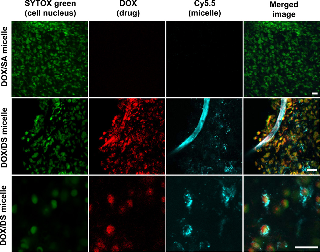
Fig. 6.
Nuclear delivery of DOX via DS micelles. Confocal fluorescence images of cancer cells in tumor tissues at 12 h after i.v. injection of DOX/SA(-Cy5.5) or DOX/DS(-Cy5.5) to M109 bearing mice. Fluorescence green, red, and cyan blue indicate SYTOX (cell nucleus), DOX (anti-cancer drug), and Cy5.5 (micelle) signals, respectively. Yellow and white colors represent the overlapping signals of DOX with SYTOX and Cy5.5. (Scale bar: 20 µm).

